Heart Anatomy

Fig 1.1 Cross-sectional view of the heart
Fig 1.2 Frontal view of the heart
The heart is a vital organ in the double, closed human circulatory system. It is a pump with 4 main chambers---- left atrium, right atrium, left ventricle, right ventricle.
The cardiac muscles formed the heart, which are the striped muscle cells, similar to skeletal cells, which contract and caused the walls to move inwards.
At the top of the heart, there is a superior (anterior) vena cava. If we look vertically downwards, we can see the inferior (posterior) vena cava. The superior vena cava transports de-oxygenated blood from the upper torso (head, body, arms) to the heart, through the 2 brachiocephalic veins. On the other hand, the inferior vena cava transport deoxygenated blood from the lower torso (abdomen, legs) to the heart. Both venae cavae carry deoxygenated blood to the right atrium and are the 2 largest veins in the body.
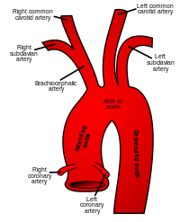
To the right of the superior vena cava, there is the aorta which is the largest artery in the heart and carries oxygenated blood away from the left atrium. In the aorta, there are 3 portions-the ascending aorta, aortic arch and descending aorta.
Fig 1.3 Aorta structure
The ascending aorta branches out into the coronary arteries. They are used to supply oxygenated blood to the heart itself, since it composes mostly of cardiac muscle tissue. The right coronary artery directs oxygenated blood to the walls of the right ventricle, right atrium, bottom portion of left ventricle and the back of septum. The left coronary artery branches again into the left anterior descending artery, which directs oxygenated blood to the left atrium, side and back of the left ventricle and the left circumflex artery which directs oxygenated blood to the front of septum, front and bottom of the left ventricle.
The aortic arch branches to the brachiocephalic artery, left common carotid artery and the left subclavian artery. The brachiocephalic artery then divides into the right common carotid artery and the right subclavian artery, as shown in Fig 1.3. The right common carotid artery extends up the right side of the neck and the left common carotid artery, to the left side. The carotid arteries supply oxygenated blood to the head and neck regions. The right subclavian artery carries oxygenated blood to the right arm and the left subclavian artery, to the left arm.
Just under the superior vena cava is the right upper heart chamber, the right atrium, which receives deoxygenated blood from the superior and inferior vena cava and pump it into the right ventricle which is then send to the lungs to be oxygenated via the pulmonary circulation The left atrium located on the left upper heart pumps oxygenated blood to the left ventricle which supplies blood to the rest of the body through the systematic circulation.
The right ventricle, below the right atrium, pumps deoxygenated blood to the pulmonary artery. The left ventricle at the lower left heart chamber, pumps oxygenated blood out at a higher pressure to the aorta.
The pulmonary arch branches out to the left and right pulmonary arteries which carry deoxygenated blood to the left and right lungs respectively. The right pulmonary artery is longer and travels the cross-section of the chest and enters the right hilum(root of the lung). The right one, fairly shorter, enters through the pericardium and into the left hilum.
The pulmonary vein, situated above the left atrium, transports oxygenated blood to the heart from the lungs, which carries oxygenated blood to the left atrium of the heart.
The median septum is a muscular wall that separates the deoxygenated blood with the oxygenated blood. Deoxygenated blood contains a higher level of carbon dioxide and a much lower ph, whereas on the other hand, oxygenated blood has a neutral ph and higher levels of oxygen. If they are mixed, it would be more difficult to rid of the acidic substances and maintaining a steady supply of oxygenated blood.
The chordae tendineae are thin fibrous threads which are attaches to one end at the edges of the tricuspid or mitral valves and another, to the papillary muscles, which are small muscles that helps to anchor these semilunar valves.
Fig1.4 Cross-section of heart showing the chordae tendineae and papillary muscles
The heart valves are categorized mainly into atrioventricular valves and semilunar valves which have a common function of preventing the blood from travelling in the backwards direction, by closing and opening due to the pressure difference. When the ventricles contract and the atrioventricular valves close, it produces a “lub” sound. When the semilunar valves close, it produces a “dub” sound. There are 4 valves in our heart chamber. The aortic and pulmonary valves belong to the semilunar valves and the tricuspid and mitral/bicuspid valves belong to the atrioventricular valves. The aortic valve is between the left ventricle and the ascending aorta, which prevents oxygenated blood from flowing back into the left ventricle. The pulmonary valve is located between the right ventricle and the pulmonary artery, preventing deoxygenated blood from flowing back into the ventricle. The tricuspid valves, located between the right atrium and the right ventricle, prevents deoxygenated blood from flowing backwards into the right atrium. The mitral valves located between the left atrium and the left ventricle, prevents oxygenated blood from flowing back to the left atrium.
The left ventricular wall is thicker than the right ventricular wall because it needs a higher pressure to pump oxygenated blood to the rest of the body, compared to the thinner right ventricular walls. which just need to pump deoxygenated blood to the lungs which is not far. Hence to handle the high amount of pressure, the left ventricular walls are thicker than the right one with a ratio proportion of 3:1.
Credits/references
http://www.medterms.com/script/main/srchcont.asp?src=cardiac+muscles&op=mm
http://biology.about.com/library/organs/heart/
http://www.texasheart.org/
http://www.cardioconsult.com/Anatomy/
http://www.mitralvalverepair.org/images/mv_anatomy/papillary-muscles.jpg
http://upload.wikimedia.org/wikipedia/commons/thumb/e/e6/Gray506.svg/200px-Gray506.svg.png
http://www.nlm.nih.gov/medlineplus/ency/images/ency/fullsize/18092.jpg
http://images.google.com/imgres?imgurl=http://www.jdaross.cwc.net/_derived/heart1.htm_txt_heart4.gif&imgrefurl=http://www.jdaross.cwc.net/heart1.htm&h=480&w=424&sz=32&hl=en&start=10&usg=__Qd0qhtdsKTZ2wgfzm2K0cfa3Xbg=&tbnid=Wvodbz0GU8goWM:&tbnh=129&tbnw=114&prev=/images%3Fq%3Dfrontal%2Bview%2Bof%2Bheart%26hl%3Den%26rlz%3D1T4SUNA_enSG257SG258
Submitted By
Hui Lin
Diwakar
Shu E
Alexis


No comments:
Post a Comment