Tissue Fluid And Blood
WHAT IS A TISSUE FLUID??
A fluid formed by the ultra filtration of blood plasma that surrounds cells. The tissue fluid acts as the internal environment between the tissue cells and the blood capillaries. It is the main component of the extracellular fluid, which also includes plasma and transcellular fluid. An analogy would be that the plasma is the “groceries”, containing useful substances, and the tissue fluid is the “trolley” which delivers the “groceries” to the tissue cells.
It mainly consist of amino acids, sugars, fatty acids, coenzymes, salts, and waste materials from cells.Its composition may depends on the exchanges between the cells and the blood.
HOW IS TISSUE FLUID FORMED?
The pumping force of the heart produces capillary hydraulic (hydrostatic) pressure, which forces water out into the interstitial space. It tries to attain equilibrium of fluid, but due to the fact that the blood is always flowing, and that the blood cells and proteins in the capillary lowers the water potential, the higher water concentration outside tends to move into the capillary via osmosis, which creates an osmotic pressure.
The one-cell thick permeable capillary allows the plasma to squeeze though the arterial walls and become tissue fluid. It also contains leucocytes, because they can change shape easily and squeeze through.
EXCHANGE OF MATERIALS BETWEEN BLOOD (IN CAPILLARIES) AND TISSUE FLUID :
The blood enters a capillary bed. These vessels are very leaky and are only wide enough for one cell at a time to pass through. The capillary walls are only one cell thick. The blood pressure forces some of the blood plasma to leak out of the capillary. This fluid is high in nutrients and oxygen (from the red blood cells). Large objects like red blood cells and protein molecules cannot pass through the walls of the capillary. The fluid that is surrounding the tissue cells is called tissue fluid. It is from this fluid that materials will diffuse into the cells. White blood cells are the only cells, which can leave the blood, so they can hunt down pathogens. Waste materials like carbon dioxide and urea diffuse from the cells into the tissue fluid. This fluid is drawn back into the blood capillary by an osmotic pressure supplied by the large proteins in the blood. Not all the tissue fluid flows back into the blood. If it did not return, the tissues would swell with fluid. Sets of vessels, called lymph vessels, drain this tissue fluid and carry it away from the tissues. Eventually the fluid (called lymph) drains back into the blood. The blood leaves the capillary beds and travels back to the heart via veins.
At the arterial end of the capillary, it faces more hydrostatic pressure than the osmotic pressure. This results in net filtration pressure towards the capillary. It would be a positive value hence fluid (plasma) flows from the capillary into the tissue fluid. This allows plasma which contains oxygen and nutrients to enter the tissue cells.
At the venous end, the net filtration pressure is a negative value as the hydrostatic pressure is less than the osmotic pressure, hence fluid moves into the capillary, to remove cellular waste products from the tissue cells.
Most of the fluid than exits the capillaries from the arterial end would enter through the venous end. The rest would be lymph, a type of interstitial fluid, which would circulate in the lymphatic system, collecting and removing the dead pathogens and cells.
http://highered.mcgraw-hill.com/sites/dl/free/0072464631/291136/Fluidexchange.swf
http://www.lymphnotes.com/article.php/id/151/
http://www.kscience.co.uk/as/module3/exercises/tissue_fluid.htm
http://en.wikipedia.org/wiki/Interstitial_fluiddone by: liting, fiona, kyaw and henry
Thursday, September 18, 2008


Monday, September 15, 2008
What is coronary heart disease?
Coronary heart disease a condition in which proper circulation of blood and oxygen are not provided to the heart and surrounding tissue. This result is due to a narrowing of the small blood vessels, which normally supply the heart with blood and oxygen.
What is angina?
Angina pectoris, commonly known as angina, is severe chest pain due to ischemia (a lack of blood and hence oxygen supply) of the heart muscle, generally due to obstruction or spasm of the coronary arteries (the heart's blood vessels). Coronary artery disease, the main cause of angina, is due to atherosclerosis of the cardiac arteries.
What is a heart attack?
A heart attack occurs when the blood supply to part of the heart is interrupted. This is most commonly due to occlusion (blockage) of a coronary artery following the rupture of a vulnerable atherosclerotic plaque, which is an unstable collection of lipids (like cholesterol) and white blood cells (especially macrophages) in the wall of an artery. The resulting ischemia (restriction in blood supply) and oxygen shortage, if left untreated for a sufficient period, can cause damage and/or death (infarction) of heart muscle tissue (myocardium).
What is atherosclerosis?
Atherosclerosis is a disease affecting arterial blood vessels. It is a chronic inflammatory response in the walls of arteries, in large part due to the accumulation of macrophage white blood cells and promoted by low density (especially small particle) lipoproteins (plasma proteins that carry cholesterol and triglycerides) without adequate removal of fats and cholesterol from the macrophages by functional high density lipoproteins (HDL), (see apoA-1 Milano). It is commonly referred to as a "hardening" or "furring" of the arteries. It is caused by the formation of multiple plaques within the arteries.

How can atherosclerosis cause heart disease?
Atherosclerosis narrows the lumen of the coronary arteries and increses heart pressure. Such an affected artery develops rough inner surfaces. This increases the risk of a blood clot being trapped in the artery. A blood clot that forms in the arteries is called thrombosis. If it occurs in t he coronary arteries, the supply of blood and oxygen to the heart muscles may be completely cut off. Without oxygen, the heart muscle cells may be damaged or die, and a heart attack occurs.
How can coronary heart disease be treated?
Occlusions in the coronary arteries can be treated with drugs and, increasingly, by a clever surgical procedure which does not involve cutting open the patient’s chest. Rather, a balloon attached to a fine wire is inserted into an artery in the leg. The balloon is then pushed through the arteries, up towards the heart until it reaches the narrow part of the coronary artery. The position of the balloon is constantly checked using X-rays. When it is in the right place, the balloon is inflated, opening up the artery and thus improving blood flow. The balloon is then pulled back out of the body.
Another way is a type of treatment called coronary bypass may also be used to improve the blood flow. This usually involves sewing a vein from the leg or artery from the chest to bypass the clogged section of an artery. The advantage is that there is no tissue rejection. Using a vein has a disadvantage in that the wall of the vein is not as thick as that of an artery, A vein also has valves, so it must be inserted in the correct direction to allow the blood to enter the heart muscles.
Resources and credits
En.wikipedia.org
Biology textbook
Done by:
Sean Tan, Soh Jun Wei, Lim Shi Zheng, Jeremy Lee
The use of CPR and detibrillation
CPR, also known as Cardio-Pulmonary Resuscitation , is a way of trying to restart the heart and breathing once they have stopped. Cardiopulmonary Resuscitation (CPR) consists of mouth-to-mouth respiration and chest compression. CPR allows oxygenated blood to circulate to vital organs such as the brain and heart. CPR can keep a person alive until more advanced procedures (such as defibrillation - an electric shock to the chest) can treat the cardiac arrest. CPR started by a bystander doubles the likelihood of survival for victims of cardiac arrest.
How to do CPR on a victim?
You will have to act quickly to help someone who may appear to have stopped breathing and has no pulse. There are a number of steps which you should follow:
Turn victim over facing up
Assess the victim’s condition - shake him gently and ask if he is all right.

Lay him face up on a firm, flat surface, moving his head and body simultaneously
Open his airway by tilting his head back and lifting the chin upwards. If you see foreign objects in his mouth or throat, remove them.
Check to see if victim is breathing
Place your face close to his mouth and observe his chest; look, listen and feel for any signs of breathing for up to 10 seconds.

If there is no indication of breathing, perform mouth-to-mouth ventilation.
Maintain an open airway. Pinch his nose and give two blows into his lungs. The duration for each breath is 1 second. The victim’s chest should rise with each blow.
Perform mouth-to-mouth ventilation
Next, feel for pulse at the carotid (neck area) for up to 10 seconds.
If there is a pulse, perform mouth-to- mouth ventilation at the rate of 12 times per minute, until natural breathing is restored.

If there is no pulse, immediately begin CPR, adhering closely to the steps that follow (Steps #6 to #7).
Centre heel one hand at lower half of victim’s breast bone keeping fingers off ribs. Keep arms straight while performing chest compressions.

If there is no pulse, perform external chest compressions as follows:
Centre the heel of one hand at the lower half of the breast bone, keeping your fingers off the ribs. Cover this hand with the heel of your other hand.
Keep your arms straight and push down vertically about 4 to 5 cm and then release. Complete 5 cycles comprising of 30 compressions & 2 full ventilations in each cycle. Between compressions, do not lift your hands off the chest.

Repeat pulse check after the 2 minute and every 5 minutes thereafter.
Stop compressions immediately once pulse returns and check for breathing
The moment his pulse returns, immediately stop compressions and check for breathing.
If victim is not breathing, perform rescue breathing at 12 times per minute ( 1 breath every 5 seconds) until victim’s natural breathing is restored.
If both pulse and breathing have returned, place victim in the recovery position and maintain an open airway.
Continue to monitor for both breathing and pulse every few minutes until help arrives.
NOTE:
To be proficient in CPR, one should undergo CPR Training to learn the proper CPR techniques and procedures.
Interesting Fact:Will CPR help someone suffering from cancer?
CPR is not always beneficial. Its success depends on many factors, including: the age and general health of the individual; any underlying health problems; the type and extent of the cancer; the main cause of the heart or breathing stopping; and how quickly the heart and breathing can be restarted.
There are over 200 different types of cancer, and many types of cancer treatment, which can affect people in very different ways. Some people may have potentially curable cancers, while others may have cancer that cannot be cured. Each individual situation is different.
If a cancer is very advanced, the heart and breathing may gradually slow down and stop as part of the natural process of dying. In this situation CPR would not be helpful or appropriate, as it is probable that the cancer itself will have affected some vital organs of the body, causing them to gradually stop working.
Sometimes a cancer may be incurable, but the person may be expected to live with the condition for a long while, sometimes years. There may be many reasons why the heart or breathing stop suddenly (such as a serious infection caused by the treatment). In this situation, attempting CPR may be appropriate, but still may not be successful.
There are four possible outcomes following cardiopulmonary resuscitation:
complete recovery partial recovery prolonged survival death. Unfortunately, only a small number of people will make a complete recovery and be discharged from hospital. Some may show a partial recovery, but will be left with serious health problems, despite every effort being made to try to help.
For some people survival may be prolonged, often by using an artificial ventilator (breathing machine) in an intensive care unit. While this may extend their life by a matter of hours, days or sometimes weeks, the person's quality of life is usually very poor. For many, CPR will not be successful and, sadly, they will still die.
Information and pictures extracted from:
http://www.scdf.gov.sg/Community_and_CD_Volunteers/Learn_Civil_Defence/First_Aid/firstaid05.html
http://depts.washington.edu/learncpr/discr.html
http://www.cancerbackup.org.uk/resourcessupport/advancedcancer/cprforpeoplewithcancer
Hope that you have learnt more about Cpr after reading our information!! Any question regarding CPR can be posted on the tag board.
Done by: Yan Ying
Sharlyn
Zarifah
Yin Kiat
Sunday, September 14, 2008
How it developed...
Our human heart is fully developed about eight weeks after conception, when the embryo is only about one inch long. The heart actually begins to beat even earlier,about four weeks after conception.
How much work is done...
Our heart pumps about 5 quarts of blood each minute which is approximately 7570 litres of blood each day throughout the body.
Do you know that your heart beats about 100,000 times each day. And doing a simple calculation, in a 70-year lifetime, the average human heart beats more than 2.5 billion times.
An adult woman's heart weighs about 8 ounces which is about 227 grams, and a man's about 10 ounces which is about 283 grams. And a child's heart is about the size of a clenched fist; an adult's heart is about the size of two fists.
It consists chiefly of muscle and can perform enough work in one hour to lift 3,000 pounds, roughly the weight of a small car, about one foot off the ground . In addition, the human heart can create enough pressure that it could squirt blood at a distance of thirty feet, amazing right?
Where is our heart...
There is common misconception that the location of our heart is at the left side of our chest.
In fact, our heart is located in the middle of your chest between your lungs. You feel more of your heart on your left side of your chest because the bottom of the heart is tipped to the left. The right lung of a human is larger than the left one. This is because of the space and placement of the heart too.
More interesting facts on blood vessels and blood....
Your system of blood vessels - arteries, veins and capillaries - is over 60,000 miles long. That's long enough to go around the world more than twice!
Do you know how often does blood go around our body?
Actually blood goes around the body about once a minute, 1500 times a day.
And an average man, has between 5 to 6 litres of blood, while an average women has between 4 to 5 litres of blood.
Do you know?
A breakup with a loved one or news of a family death literally can lead to broken hearts in the form of heightened risk for heart attack, studies have shown. Such trauma can also trigger the release of stress hormones into the bloodstream that temporarily "stun" the heart.
People say "Bless you" when you sneeze because when you sneeze, your heart stops for a millisecond.
Women hearts beat faster than men. The main reason for this is simply that on average women tend to be smaller than men and have less mass to pump blood to.
Resources and credits:
http://my.clevelandclinic.org/heart/heartworks/heartfacts.aspx
http://www.stunning-stuff.com/list-funny-facts/5.html?pg=5
http://www.funshun.com/amazing-facts/heart-human-body-facts.html
http://www.muskurahat.com/amazing-facts/Heart-facts-2.asp
http://wiki.answers.com/Q/What_are_interesting_facts_about_the_heart
Book : Human body, written by Angela Royston
Illustrated by Mike Suanders
Lastly, this is a video to help us understand heart diseases better.
Done By:
XingJie
LooPin
JingWen
Qinghua
the BLOOD group

The Blood Group------------------Experiments with blood transfusions, the transfer of blood or blood components into a person's blood stream, have been carried out for hundreds of years. Many patients have died and it was not until 1901, when the Austrian Karl Landsteiner discovered human blood groups, that blood transfusions became safer.Mixing blood from two individuals can lead to blood clumping or agglutination. The clumped red cells can crack and cause toxic reactions. This can have fatal consequences. Karl Landsteiner discovered that blood clumping was an immunological reaction which occurs when the receiver of a blood transfusion has antibodies against the donor blood cells.Karl Landsteiner's work made it possible to determine blood types and thus paved the way for blood transfusions to be carried out safely. For this discovery he was awarded the Nobel Prize in Physiology or Medicine in 1930
All humans and many other primates can be typed for the ABO blood group.Basically,there are four principal types: A, B, AB, and O. There are two antigens and two antibodies that are mostly responsible for the ABO types.Very often,an individual’s type is determined by the specific combination of these four components. Individuals with type O blood do not produce ABO antigens. Therefore, their blood normally will not be rejected when it is given to others with different ABO types.This results that type O people are normally named as universal donors for transfusions.Despite being an universal donors,they can receive only type O blood themselves.
Another exception is that those who have type AB blood do not make any ABO antibodies.Hence,Their blood does not discriminate against any other ABO type. Consequently, they are universal receivers for transfusions, but their blood will be agglutinated when given to people with every other type because they produce both kinds of antigens.

By a few drops of blood,you could determine an individual’s ABO type.
Mixed some of the blood with a serum containing anti-A antibodies. Another serum with anti-B antibodies is mixed with the remaining sample.Based on theory of blood group,whether or not agglutination will occur in either sample indicating the ABO type. It is a simple process of elimination of the possibilities. For instance, if an individual's blood sample is agglutinated by the anti-A antibody, but not the anti-B antibody, it means that the A antigen is present but not the B antigen. Therefore, the blood type is A.
Special Thanks to :
http://nobelprize.org/educational_games/medicine/landsteiner/readmore.html
http://anthro.palomar.edu/blood/ABO_system.htm
submitted by:
vanessa
pamelyn
xueting
wenxiu
:)
Therefore, it refers to the force exerted by the blood circulation on the walls of the blood pressure.
How is blood pressure measured?
- maximum pressure in an artery at the moment when the heart is beating and pumping blood through the body.
2) Diastolic Pressure
- lowest pressure in an artery in the moments between beats when the heart is resting.
2) Another tube leads from the cuff to a reservoir of mercury at the bottom of a vertical glass column. Whatever pressure is in the cuff is shown on the mercury column. The mercury is held within a sealed system – only air travels in the rubber tubing and the cuff.
3) Air is then blown into the cuff and increasing pressure and tightening is felt on the upper arm.
4) The doctor puts a stethoscope to your arm and listens to the pulse while the air is slowly let out again.
5) The systolic pressure is measured when the doctor first hears the pulse.
6) This sound will slowly become more distant and finally disappear.
7) The diastolic pressure is measured from the moment the doctor is unable to hear the sound of the pulse.
Stethoscope & Sphygmomanometer
Below is a video of how to measure blood pressure:
2) The pressure in the left ventricle becomes higher than that in the aorta. The semi-lunar valve in the aorta opens.
3) The ventricle begins to relax. The aortic valve closes to prevent backflow of blood into the ventricle.
4) The pressure in the ventricle continues to decrease as it relaxes.
5) The bicuspid valve opens as the pressure in the ventricle becomes lower than that in the atrium.
6) The pressure in the ventricle gradually increases as blood continues to enter the ventricle fromt the atrium
7) The cycle repeats.
How does blood pressure differ in differetn blood vessels?
Each blood vessel has it's own blood pressure value, arterial blood pressure, capillary blood pressure, venous blood pressure, left atrial blood pressure, right ventricular blood pressure etc.


THE CARDIAC CYCLE.
Diastole - Ventricles are relaxed.
Systole - Ventricles contract.
During the diastole phase the atria and ventricles are relaxed and the atrioventricular valves are open. De-oxygenated blood from the superior and inferior vena cava flows into the right atrium. The open atrioventricular valves allow blood to pass through to the ventricles. The SA* node contracts triggering the atria to contract. The right atrium empties its contents into the right ventricle. The tricuspid valve prevents the blood from flowing back into the right atrium.
During the systole phase the right ventricle receives impulses from the Purkinje fibers and contracts. The atrioventricular valves close and the semi lunar valves open. The de-oxygenated blood is pumped into the pulmonary artery. The pulmonary valve prevents the blood from flowing back into the right ventricle.
The pulmonary artery carries the blood to the lungs. There the blood picks up oxygen and is returned to the left atrium of the heart by the pulmonary veins.
In the next diastole period, the semi lunar valves close and the atrioventricular valves open. Blood from the pulmonary veins fills the left atrium. (Blood from the vena cava is also filling the right atrium.) The SA node contracts again triggering the atria to contract. The left atrium empties its contents into the left ventricle. The mitral valve prevents the oxygenated blood from flowing back into the left atrium.
During the systole phase the atrioventricular valves close and the semi lunar valves open. The left ventricle receives impulses from the Purkinje fibers and contracts. Oxygenated blood is pumped into the aorta. The aortic valve prevents the oxygenated blood from flowing back into the left ventricle.
The aorta branches out to provide oxygenated blood to all parts of the body. The oxygen depleted blood is returned to the heart via the vena cava.
*SA node is the Sinoatrial Node which is also refer as the pacemaker of the heart as it set the contraction of the heart.
click on the links for Videos:
http://www.youtube.com/watch?v=sRJHn7ctBCo&feature=related
http://www.youtube.com/watch?v=H04d3rJCLCEhttp://www.youtube.com/watch?v=4ZzuqBM8nPU&NR=1
references:
http://images.google.com.sg/imgres?imgurl=http://z.about.com/d/biology/1/0/i/1/cycle.gif&imgrefurl=http://biology.about.com/b/2006/01/14/cardiac-cycle-diastole-phase.htm&h=291&w=301&sz=10&hl=en&start=8&usg=__73ABG3ms2FOkrSBZk4F57c_B068=&tbnid=XjbR6xdNNp2vDM:&tbnh=112&tbnw=116&prev=/images%3Fq%3Dcardiac%2Bcycle%26gbv%3D2%26hl%3Den

Picture taken from http://intensivecare.hsnet.nsw.gov.au/five/images/heart6.jpg

done by: jyun yao
felicia
weikang
norman
Saturday, September 13, 2008
Smoking, Diet and the heart
Smoking
Smoking can be equally harmful and deadly to the heart as taking drugs such as heroin and cocaine. It is is a major cause of heart attack, stroke and peripheral arterial disease. Not only smokers are affected; passive smokers are too.
Each cigarette contains over 4,000 harmful chemicals, of which 400 are poisonous and 50 can cause cancer.
These include:
· TAR
· CARBON MONOXIDE
· NICOTINE
· DDT
· ARSENIC
· AMMONIA
And these are just 6 out of the 400.
Now let's look at some harms that smoking does to the heart.
Coronary Heart Disease

Angina
Angina or "angina pectoris," is also known as chest pain. Angina can be described as a discomfort, heaviness, pressure, aching, burning, fullness, squeezing or painful feeling. It is usually felt in the chest, but may also be felt in the shoulders, arms, neck, throat, jaw or back.

What causes Angina?
When blood flow to an area of the heart is decreased, it impairs the delivery of oxygen and vital nutrients to the heart muscle cells. When this happens, the heart muscle must use alternative, less efficient forms of fuel so that it can perform its function of pumping blood to the body. The byproduct of using this less efficient fuel is a compound called lactic acid that builds up in the muscle and causes pain.
Smoking causes a fatty build-up in the blood vessels. These fatty deposits result in narrowing and blockage of the blood vessels.
The moment you light up your cigarette, thousands of poisonous chemicals in the cigarette smoke enter your body and get absorbed into the bloodstream. Some of them damage the lining of the blood vessels by making the blood vessel walls sticky and hence causing them to collect tiny fatty deposits that float in the bloodstream. The more you smoke, the more the deposits build up.
Similar adverse effects take place when the blood vessels leading to our heart are blocked by fatty deposits. This cuts off the blood supply to the heart muscles. As a result, the tissue in that area will die and hence, a heart attack occurs.


-Non-smokers living with smokers have about a 25 to 30 per cent increase in risk of heart disease and are also more likely to suffer a stroke.
-Exposure to secondhand smoke is especially risky for children and babies and may cause low birth weight, sudden infant death syndrome (SIDS), bronchitis, pneumonia, asthma and middle ear infections.
So in order to prevent coronary heart diseases, we must not smoke. By smoking, it increases your risk of heart attack by two to six times! Also, we must take extra care of our diet. How to? Avoid consuming too much fats, protein and carbohydrate.
FATS
Avoid eating too much Trans fat and saturated fat.
Trans fat are artery-clogging element of partially hydrogenated oils. They are found in many fried foods and commercial baked goods. These fats increase your bad cholesterol (LDL) while lowering your good cholesterol (HDL) hence, weakening your natural defences against heart disease. Harvard researchers have estimated that banning Trans fats from the American diet could prevent some 228,000 heart attacks each year.
Saturated fat in meat or dairy products encourages a buildup of cholestrol in the arteries by increasing LDL levels in the blood.
Even though some fats are unhealthy to our body, some also helps, like Unsaturated fats and omega-3 fatty acids. Unsaturated fat are found in olive oil, canola oil, nuts, avocados, and fish can actually clear LDL while boosting HDL.
Omega-3 fatty acids are found in cold-water fish. It has been shown to prevent blood clots, lower blood pressure and slow the buildup of cholestrol in the arteries. Large studies suggest that this fat can lower the risk of heart disease by more than 35% and can reduce the risk of sudden death from heart attack by more than 50%.
PROTEIN
Medical research shows that consuming too much protein, more than 30% of your total daily caloric intake could actually harm your body.
A diet in which protein makes up more than 30% of your caloric intake causes a buildup of toxic ketones (waste products when your body burns stored fat for energy). Ketogenic diets can push your kidneys into overdrive in order to flush these ketones from your body. As your kidneys rid your body of these toxic ketones, you can lose a significant amount of water, which puts you at risk of dehydration, especially if you exercise heavily.
The water loss often shows up as weight loss. But along with losing water, you lose muscle mass and bone calcium. The dehydration also strains your kidneys and puts stress on your heart.
CARBOHYDRATES
The total storage capacity carbohydrate in our body is really quite limited. An average person can store about three hundred to four hundred grams of carbohydrate in his muscles but these carbohydrates are not of use. In the liver, where carbohydrates are accessible for glucose conversion, you can store only about sixty to ninety grams. This is equivalent to about two cups of cooked pasta or three typical candy bars, and it represents your total reserve capacity to keep the brain working properly.
It was thought that an increase in fat would lead to high cholesterol which is associated with heart disease. When the studies were actually done, the opposite happened. People on a low-carbohydrate diet improved their cholesterol readings even when they increased their fat intake and even when their intake of saturated fat increased. It appears that your body must rely on fat for energy and the saturated fat you eat gets burned up before it can cause any harm.
Acknowledgements
http://www.betterhealth.vic.gov.au/bhcv2/bhcarticles.nsf/pages/Smoking_and_heart_disease_the_facts http://www.sads.org.uk/heart_function.htm
http://www.webmd.com/heart-disease/guide/heart-disease-angina
http://www.heart.co.il/images/img_01_Education/angina.jpg
http://www.hpb.gov.sg/hpb/default.asp?pg_id=1238&altid=0
http://www.hpb.gov.sg/hpb/default.asp?pg_id=865&aid=113
http://www.hpb.gov.sg/hpb/default.asp?pg_id=865&aid=106
http://www.hpb.gov.sg/hpb/default.asp?pg_id=865&aid=106&altid=0
http://rheumatic.org/insulin.htm
Most of the above information is abstracted from books by Dr. Sears: "Enter the Zone", and Dr. Maffetone "In Fitness and in Health".
http://www.drjaywortman.com/blog/wordpress/my-big-fat-diet-faqs/
http://www.medicinenet.com/script/main/art.asp?articlekey=50900
http://www.abbottdiabetescare.com/adc_dotcom/url/questionAnswerProfile/en_US/40.20:20/question_answer/question_answer/QuestionAnswer_00146.htm http://www.health.com/health/condition-article/0,,20189107,00.html
http://www.youtube.com/watch?v=GBf59Z8tgA0
‘A HEALTH GUIDE FOR SCOUTS’
Report written by:
Chia Kiah Yeen
Michelle
Wong Jyun Yir
Neo Yao Kian Andy
Heart Anatomy
Heart Anatomy

Fig 1.1 Cross-sectional view of the heart
Fig 1.2 Frontal view of the heart
The heart is a vital organ in the double, closed human circulatory system. It is a pump with 4 main chambers---- left atrium, right atrium, left ventricle, right ventricle.
The cardiac muscles formed the heart, which are the striped muscle cells, similar to skeletal cells, which contract and caused the walls to move inwards.
At the top of the heart, there is a superior (anterior) vena cava. If we look vertically downwards, we can see the inferior (posterior) vena cava. The superior vena cava transports de-oxygenated blood from the upper torso (head, body, arms) to the heart, through the 2 brachiocephalic veins. On the other hand, the inferior vena cava transport deoxygenated blood from the lower torso (abdomen, legs) to the heart. Both venae cavae carry deoxygenated blood to the right atrium and are the 2 largest veins in the body.
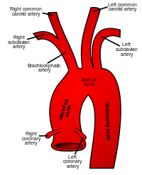
To the right of the superior vena cava, there is the aorta which is the largest artery in the heart and carries oxygenated blood away from the left atrium. In the aorta, there are 3 portions-the ascending aorta, aortic arch and descending aorta.
Fig 1.3 Aorta structure
The ascending aorta branches out into the coronary arteries. They are used to supply oxygenated blood to the heart itself, since it composes mostly of cardiac muscle tissue. The right coronary artery directs oxygenated blood to the walls of the right ventricle, right atrium, bottom portion of left ventricle and the back of septum. The left coronary artery branches again into the left anterior descending artery, which directs oxygenated blood to the left atrium, side and back of the left ventricle and the left circumflex artery which directs oxygenated blood to the front of septum, front and bottom of the left ventricle.
The aortic arch branches to the brachiocephalic artery, left common carotid artery and the left subclavian artery. The brachiocephalic artery then divides into the right common carotid artery and the right subclavian artery, as shown in Fig 1.3. The right common carotid artery extends up the right side of the neck and the left common carotid artery, to the left side. The carotid arteries supply oxygenated blood to the head and neck regions. The right subclavian artery carries oxygenated blood to the right arm and the left subclavian artery, to the left arm.
Just under the superior vena cava is the right upper heart chamber, the right atrium, which receives deoxygenated blood from the superior and inferior vena cava and pump it into the right ventricle which is then send to the lungs to be oxygenated via the pulmonary circulation The left atrium located on the left upper heart pumps oxygenated blood to the left ventricle which supplies blood to the rest of the body through the systematic circulation.
The right ventricle, below the right atrium, pumps deoxygenated blood to the pulmonary artery. The left ventricle at the lower left heart chamber, pumps oxygenated blood out at a higher pressure to the aorta.
The pulmonary arch branches out to the left and right pulmonary arteries which carry deoxygenated blood to the left and right lungs respectively. The right pulmonary artery is longer and travels the cross-section of the chest and enters the right hilum(root of the lung). The right one, fairly shorter, enters through the pericardium and into the left hilum.
The pulmonary vein, situated above the left atrium, transports oxygenated blood to the heart from the lungs, which carries oxygenated blood to the left atrium of the heart.
The median septum is a muscular wall that separates the deoxygenated blood with the oxygenated blood. Deoxygenated blood contains a higher level of carbon dioxide and a much lower ph, whereas on the other hand, oxygenated blood has a neutral ph and higher levels of oxygen. If they are mixed, it would be more difficult to rid of the acidic substances and maintaining a steady supply of oxygenated blood.
The chordae tendineae are thin fibrous threads which are attaches to one end at the edges of the tricuspid or mitral valves and another, to the papillary muscles, which are small muscles that helps to anchor these semilunar valves.
Fig1.4 Cross-section of heart showing the chordae tendineae and papillary muscles
The heart valves are categorized mainly into atrioventricular valves and semilunar valves which have a common function of preventing the blood from travelling in the backwards direction, by closing and opening due to the pressure difference. When the ventricles contract and the atrioventricular valves close, it produces a “lub” sound. When the semilunar valves close, it produces a “dub” sound. There are 4 valves in our heart chamber. The aortic and pulmonary valves belong to the semilunar valves and the tricuspid and mitral/bicuspid valves belong to the atrioventricular valves. The aortic valve is between the left ventricle and the ascending aorta, which prevents oxygenated blood from flowing back into the left ventricle. The pulmonary valve is located between the right ventricle and the pulmonary artery, preventing deoxygenated blood from flowing back into the ventricle. The tricuspid valves, located between the right atrium and the right ventricle, prevents deoxygenated blood from flowing backwards into the right atrium. The mitral valves located between the left atrium and the left ventricle, prevents oxygenated blood from flowing back to the left atrium.
The left ventricular wall is thicker than the right ventricular wall because it needs a higher pressure to pump oxygenated blood to the rest of the body, compared to the thinner right ventricular walls. which just need to pump deoxygenated blood to the lungs which is not far. Hence to handle the high amount of pressure, the left ventricular walls are thicker than the right one with a ratio proportion of 3:1.
Credits/references
http://www.medterms.com/script/main/srchcont.asp?src=cardiac+muscles&op=mm
http://biology.about.com/library/organs/heart/
http://www.texasheart.org/
http://www.cardioconsult.com/Anatomy/
http://www.mitralvalverepair.org/images/mv_anatomy/papillary-muscles.jpg
http://upload.wikimedia.org/wikipedia/commons/thumb/e/e6/Gray506.svg/200px-Gray506.svg.png
http://www.nlm.nih.gov/medlineplus/ency/images/ency/fullsize/18092.jpg
http://images.google.com/imgres?imgurl=http://www.jdaross.cwc.net/_derived/heart1.htm_txt_heart4.gif&imgrefurl=http://www.jdaross.cwc.net/heart1.htm&h=480&w=424&sz=32&hl=en&start=10&usg=__Qd0qhtdsKTZ2wgfzm2K0cfa3Xbg=&tbnid=Wvodbz0GU8goWM:&tbnh=129&tbnw=114&prev=/images%3Fq%3Dfrontal%2Bview%2Bof%2Bheart%26hl%3Den%26rlz%3D1T4SUNA_enSG257SG258

Submitted By
Hui Lin
Diwakar
Shu E
Alexis
Tuesday, September 9, 2008
Of Veins and Valves
Veins in the human body carry deoxygenated blood towards the heart, with one exception: the pulmonary veins. These veins carry oxygenated blood from the lungs to the heart.
The blood in veins flows slower and smoother than in arteries and the pressure, too, is much lower. Because of this, the walls of the veins contain less elastic fibres and muscles than in the walls of the arteries. The picture below shows a photomicrograph of the cross sections of an artery and a vein.

Picture taken from www.spjc.edu/clw/math_science/Nicotera/pnic/Nicotera/artery%20and%20vein%201.jpg

Skeletal muscles and veins
Due to the low pressure and slow flow of blood in veins, skeletal muscles that surround these veins help in moving the blood along. When skeletal muscles contract, they compress the veins, forcing blood to move along more quickly.
Role of valves in veins
Most veins contain a special feature which other blood vessels do not have. They contain valves. Valves prevent the backflow of blood and this is important in veins because as blood flows to the heart, it may momentarily be pulled backwards due to gravity or muscle contraction. These valves would then immediately close to prevent blood from flowing back. The presence of valves also ensures that blood flows in one direction only, which is, back to the heart.

Picture taken from www.merck.com/mmhe/sec03/ch036/ch036a.html
For an animation on this, click here. Observe the blood cells and see how the valves close when blood moves backwards momentarily.
Characteristic of venous valves
Venous valves are folds on the lining layer of a vein, shaped like half-moons.
Lastly, here's a short video clip taken from youtube, which compares the structures of arteries and veins, and discusses how blood moves along in veins.
References:
- http://www.youtube.com/
- The Merck Manuals: Online Medical Library
www.merck.com/mmhe/sec03/ch036/ch036a.html - Dr. Phil Nicotera's site at St. Petersburg College
www.spjc.edu/clw/math_science/Nicotera/pnic/Nicotera/artery%20and%20vein%201.jpg - The Doe Report by Medical Legal Art
http://www.doereport.com/generateexhibit.php?ID=14555&ExhibitKeywordsRaw=&TL=4294967295&A - Centre for Cancer Foundation
http://cancerweb.ncl.ac.uk/cgi-bin/omd?venous+valve
Written by Mrs Eunice Chan